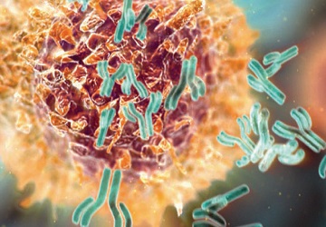
The main goal of our research is to define the regulation of adaptive immune responses during inflammation within the central nervous system (CNS). The Wu lab has several areas of ongoing investigation into the pathogenesis of multiple sclerosis (MS) and related diseases. We are exploring various immunologic abnormalities in MS by examining characteristics of circulating B cells and monocytes from patients. In addition, we are modeling cellular and molecular immune contributions to neuro-inflammation by primarily utilizing murine experimental autoimmune encephalomyelitis (EAE). Finally, we are actively engaged in several clinical trials to identify potential new highly efficacious disease-modifying therapies and mechanisms of action for currently approved MS therapies.
This is for making space so that the pictures aren't so close to the text. I wish I knew how to make it so that I can make a space without doing this. Also if I don't do this there is a little bit of publication that pops up at the bottom of the page so I'm gonna do this so that it doesn't do that.


This is for spacing
Our understanding of the basic pathogenic mechanisms of MS has been radically changed by the emergence of novel immune-based treatments that have specifically targeted B cells. Because there is strong clinical and experimental evidence that B cells contribute to MS via their role as antigen presenting cells, we have used our conditional mouse system to explore the extent to which B cells alone can drive CD4 T cell auto-reactivity in vivo. In our preliminary studies, we have found that antigen presentation by B cells alone is not sufficient to support passive EAE resulting from transfer of encephalitogenic CD4 T cells, unless B cells also express antigen receptor specific for cognate antigen. Currently, we are exploring the mechanisms by which B cells drive CD4 T cell auto-reactivity during EAE and the role of B cell antigen presentation in meningeal ectopic lymphoid tissue development.
Spacing
This is for spacing
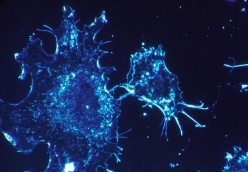
The goal of this project is to identify how immune cells access immune targets that are generated within the CNS. Acquisition and processing of myelin targets are critical to the initiation and propagation of autoimmune CNS inflammation. How antigens from within the compartmentalized CNS are accessed by APCs has yet to be established. We hypothesize that various CNS targets are available to APCs such as dendritic cells and B cells within secondary lymphoid tissue.
In collaboration with Dr. Mark Miller, we are using two-photon microscopy (TPM) to test this hypothesis. We are examining the behavior of myelin-oligodendrocyte glycoprotein-specific CD4 T cells before and after the development of neuro-inflammation in EAE. Experiments will test the capacity for different APC subsets to capture and present antigen to autoreactive CD4 T cells as well as determine the potential availability of myelin antigens in the periphery of patients with MS.
Spacing
This is for spacing
Myelin is targeted in MS by both innate and adaptive immune cells. We have observed a role for a vanilloid-type member of the Transient Receptor Potential (TRP) channel family, TRPV4 in EAE. In collaboration with Hongzhen Hu from the Center for the Study of Itch, we have observed in preliminary studies the expression of TRPV4 by immune cells. We are presently testing the requirement for innate immune cell expression of TRPV4 during neuro-inflammation using in vivo genetic manipulation of TRPV4. Additionally, we are pursuing studies using human specimens to explore whether TRPV4 is involved in the development of inflammatory demyelinating MS plaques.
Spacing
This is for spacing
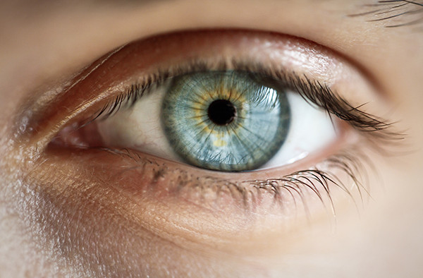
In collaboration with Dr. Beau Ances and Dr. Greg Van Stavern, we have piloted a study to examine the cerebral changes resulting from anterior visual pathway loss in patients with optic neuritis. We are seeking to understand how the process of neuro-inflammation in patients with early forms of MS involving the optic nerve affects neuronal activity within visual pathways. We have utilized a new MRI technique, functional connectivity (fcMRI), to assess neuronal function in the visual system in patients who have suffered from inflammatory damage to the anterior visual pathway.
The objective of this study is to determine whether the visual pathways in the brain are altered in the earliest stages of MS. Patients with optic neuritis have undergone evaluation with clinical measures, including vision tests, optical coherence tomography and visual function questionnaires; along with imaging of the brain using fcMRI and standard MRI sequences.
In collaboration with Dr. Joe Culver, we are also pursuing correlates of this imaging technique in animal models of optic neuritis. Ultimately, assessment by this form of imaging may be useful in determining treatment response and severity of disease in patients with inflammatory demyelinating disease.
Spacing
This is for spacing
CLOCK-MS is a multi-site, phase IV clinical trial (sponsored by EMD Serono) designed to understand the mechanism of action of Cladribine (Mavenclad®) tablets by exploring the effect on CNS biomarkers relevant in the relapsing forms of MS. The study is designed to generate hypotheses regarding the impact and relevance of Cladribine tablet activity in the CNS by assessing the cerebrospinal fluid (CSF) levels of lymphocyte subsets, other immune cells, neuronal injury markers, and soluble immunological markers in study participants with relapsing forms of MS before and during treatment with Cladribine, and the association of these CSF markers with corresponding blood markers and with clinical outcomes. ClinicalTrials.gov Identifier: NCT03963375
Spacing